Lacrimal Duct Surgery
The Lacrimal Duct Surgery is often required when Lacrimal Duct Dilatation procedure showed the absence of the drain tube or complete closure due to scarring from infection. This procedure is often called Dacryocystorhynostomy (DCR) which is creating a new tear drain.
What is DCR?
DCR is a surgery performed to creat a new tear drain between the eye and nose when your current tear drain becomes blocked or obstructed.
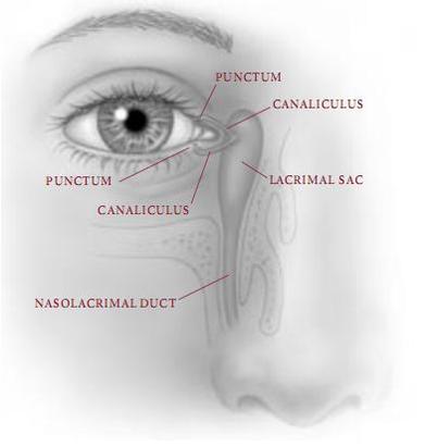
What is the anatomy?
The tear drain consists of two small openings called punctum; one in your upper eyelid and the other in your lower eyelid. Each of these openings leads into a small tube called the canaliculus which, in turn, empties into the lacrimal sac between the inside corner of your eye and your nose. The lacrimal sac leads into a canal called the nasolacrimal duct that passes through the bony structures surrounding your nose and empties tears into your nasal cavity.
How does the tear drain work?
When you blink, your eyelids push tears evenly across the eyes to keep them moist and healthy. Blinking also pumps your old tears into the punta and lacrimal sac where they travel through the tear duct and drain into your nose. If the tear duct is blocked, your tears back up and spill over your eyelids as if you were crying. Tears trapped in the tear sac also can become stagnant and infected. A DCR can be performed to correct this problem.
What are the symptoms of having a plugged and infected tear drain?
The most common symptoms are excessive watering, mucous discharge, eye irritation, and painful swelling in the inner corner of your eyelids. A skillful history and physical examination can usually pinpoint the cause of tearing. If your symptopms go untreated, an infection can develop around your eye.
What are the treatments?
Your doctor may recommend a number of treatments based on the analysis of your symptoms. In some instances, it may be as simple as applying warm compresses and antibiotics. Usually, non-invasive approaches (e.g. Lacrimal Duct flushing, Lacrimal Duct Dilatation etc) will be administered first and surgery will be the last option if all else fail.
In the case where surgery is required, your surgeon will create a new tear drain opening from your blocked sac directly into your nose to bypass the obstruction. A small incision is made either in the skin or inside the nose. A fine, soft silicone stent may temporary be left in the new tear drain (for between one to six months after surgery) to keep the duct open while healing occurs. If the obstruction cannot be opened, it may necessary to surgically place a tiny artificial drain called a Jones Tube behind the inner corner of the eyelids. The tube is made of Pyrex glass and remains permanently in the tear duct.
What are the risks and complications?
In addition to the removal of the sutures, minor bruising or swelling may be expected and will likely go away in one or two weeks. Occasionally, scar tissue may form, blocking the drain again, which may require Lacrimal Duct Dilatation or repeating the surgical procedure.
Bleeding and infection are potential risks with any surgery. Hence, one should always consider non-invasive approach first.
Is the surgery effective?
Most patients experience resolution of their tearing and discharge once surgery is completed, with little, if any, postoperative discomfort.
Who performs the surgery?
Patients are most commonly treated by opthalmic plastic and reconstructive surgeons who specialize in diseases and problems of the eyelids, tear drain, and orbit (the area around the eye).